Contact us
Call us today to schedule an appointment or to learn more about our Hip Preservation Program.
A PAO is a surgery used to correct a condition called hip dysplasia or acetabular dysplasia. Hip dysplasia is when there is insufficient coverage of the femoral head (ball) by an excessively shallow acetabulum (hip socket). During a PAO, the acetabulum is repositioned to cover more of the femoral head to improve the stability of your child’s hip joint. The PAO surgery improves your child’s hip function, decreases hip pain, and stops the damage occurring inside the joint that can lead to hip arthritis over time.
The PAO preserves the integrity of the pelvic ring but allows precise and full correction of even severe hip dysplasia. It involves cutting the pelvis around the entire acetabulum, which is then moved into a position that better covers the femoral head. Usually, three or four screws are used to hold the acetabulum in its new position. Over time, new bone will grow where the cuts were made, fusing the acetabulum to the rest of the pelvis.
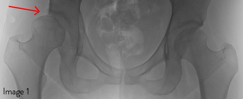
Image 1: The red arrow points to the dysplasia of the right hip. Compared with the normal left hip, the right femoral head is not covered by the acetabulum and is starting to slip upward and out of the socket.
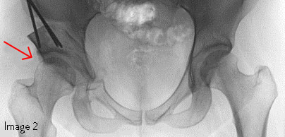
Image 2: The red arrow points to the right hip after a PAO has been performed. The femoral head is now properly covered and has moved downward and into the acetabulum.
Occasionally, the hip joint may need to be opened or a hip arthroscopy may need to be performed at the same time as a PAO to repair damage inside the joint, such as a labral tear.
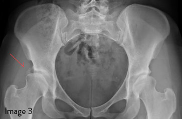
Image 3: The red arrow points to the dysplasia of the right hip. Notice the decreased lateral coverage of the right femoral head by the acetabulum compared with the coverage of the left femoral head.
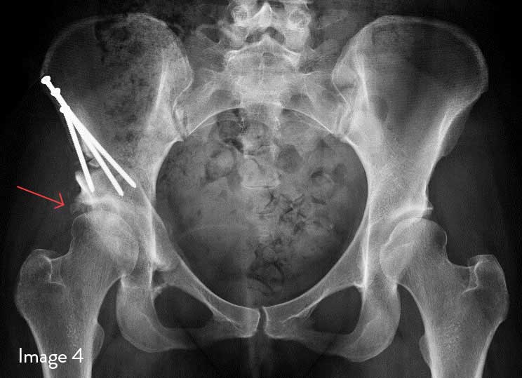
Image 4: The red arrow points to the right hip after a PAO has been performed. The femoral head is now properly covered.
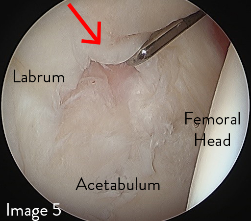
Image 5: During the PAO surgery, a hip arthroscopy was performed, allowing visualization of the cartilage and labrum. The red arrow points to a labral tear.
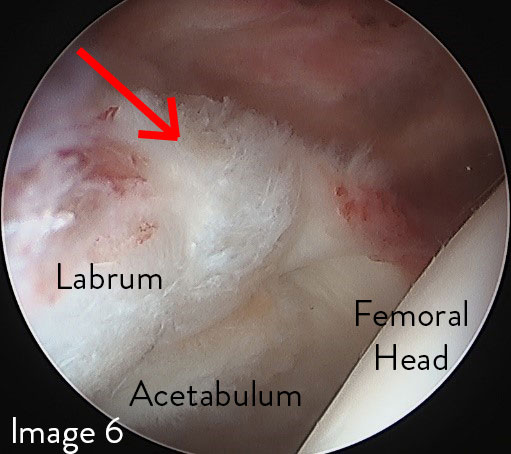
Image 6: The red arrow points to where the labral tear was repaired. If femoral deformity is also contributing to your child’s hip dysplasia, then a PAO may be performed in conjunction with a femoral osteotomy. Usually, the femoral osteotomy is performed through a second incision over the side of the thigh.
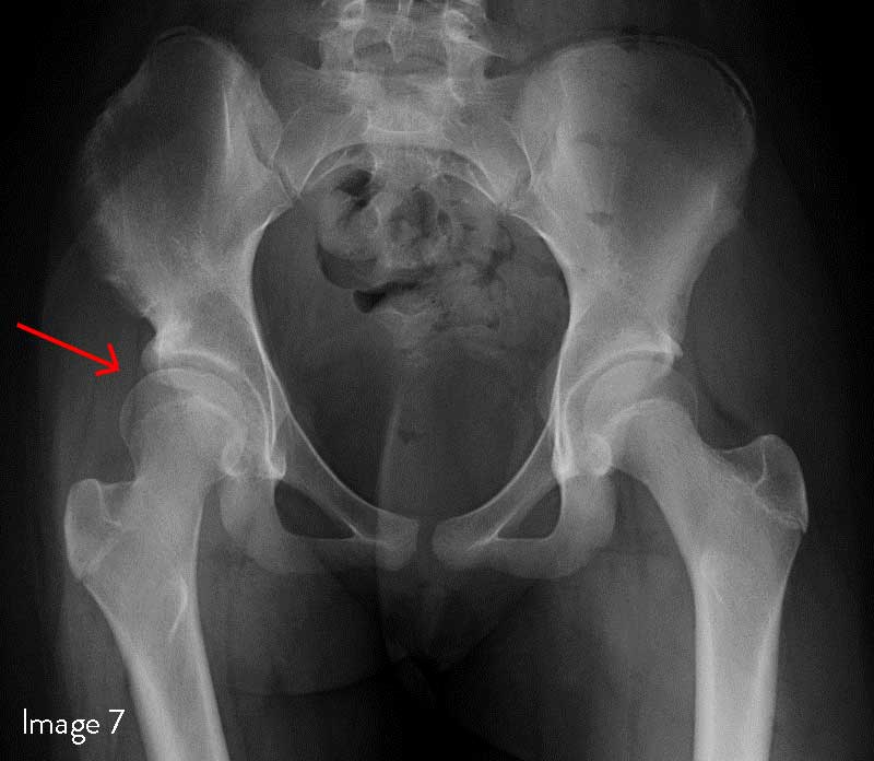
Image 7: The red arrow points to the dysplasia of the right hip. Compared with the normal left hip, the right femoral head is not covered by the acetabulum, and the femoral head is pointing excessively upward.
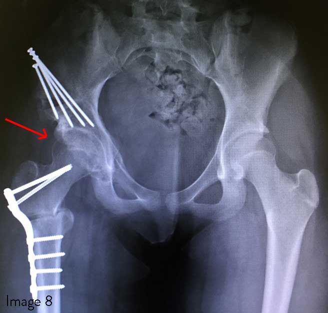
Image 8: The red arrow points to the right hip after a PAO combined with a femoral osteotomy has been performed. The femoral head is now properly covered and pointing toward the center of the socket. The right hip appears more symmetrical with the normal left hip.
After a PAO, you can expect your child to be:
Most patients are completely healed and back to sports three to six months after surgery.
Patients have reduced pain, improved hip function, and increased activity-level scores in short-term-outcome studies two years after a PAO. The 10-year outcomes for the PAO procedure show that 80% to 90% of patients are free of end-stage osteoarthritis.
Call us today to schedule an appointment or to learn more about our Hip Preservation Program.

Connect with us:
Download our App: