Contact us
Refer a patient. For more information, please call
We use state-of-the-art imaging technology and techniques to capture images with a level of detail, clarity and speed never before possible. These images ultimately help our radiologists quickly and accurately diagnose your child’s condition.
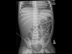
X-ray - An x-ray takes a picture of the bones inside your child’s body. Each picture takes just a couple of seconds, like a regular photograph.
Computed tomography (CT) - A CT scanner is an x-ray camera that very quickly takes multiple pictures of the inside of your child’s body and carefully adds the pictures together to make a 3-D picture. Compared to older CT scanners, our high-speed CT scanner allows us to take very sharp images. It’s also better at managing small movements a child may make during an exam.

Magnetic resonance imaging (MRI) - The MRI machine uses magnets, radio waves and a computer to take very detailed pictures of the inside of your child’s body. Your child will have to stay very still for about 30 to 60 minutes during the MRI. Some children may receive anesthesia to help them stay still. Compared to older MRI machines, our 3T MRI machine and advanced processing protocols provide more-detailed images in less time.

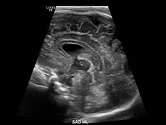
Ultrasound - Ultrasounds use high-frequency sound waves to create an image of the inside of your child’s body.
Fluoroscopy - Fluoroscopy is a continuous, movie-like x-ray. It shows how a body part moves or how something moves through the body.
PET/MRI - PET/MRI combines images from two different technologies, allowing us to see the form and function of organs and tissues while reducing radiation exposure.
SPECT/CT - This test also combines two different technologies and enables us to see both the 3-D form of the part of the body under study and its function at the same time. Combining SPECT and CT imaging in one test reduces the number of doctor’s office visits our patients have to make and help us make more accurate diagnoses.
Bone density or DEXA - This test measures bone strength and can track changes over time.
Contrast. Some procedures may require your child to take contrast. Contrast is a fluid that your child will drink or get through an IV (or both). Once it is inside your child’s body, it looks extra bright in radiology images and can make it easier for the doctors to see what they’re looking for.
Our pediatric radiologists have completed specialized training to take and interpret images of a child’s:
Learn more about our team devoted to using imaging to diagnose conditions in utero during pregnancy.
Refer a patient. For more information, please call

Connect with us:
Download our App: