Call us today
Contact us to learn more about our pediatric Chest Wall Program or to schedule an appointment.
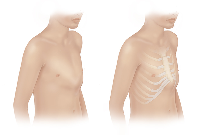
Pectus carinatum, also called "pigeon chest," is a deformity of the chest characterized by a protrusion of the sternum (breastbone) and ribs. It occurs more often in boys and typically becomes more pronounced during early adolescence.
At the Stanford Medicine Children’s Health Chest Wall Clinic, your child will meet with our team as we obtain a complete history and physical exam. We will also perform 3D mapping of your child’s chest wall during the clinic appointment. We may recommend additional diagnostic testing.
At Stanford Medicine Children’s Health, we continually stay on the leading edge of treatment options for chest wall deformities. For pectus carinatum, we offer two types of treatment: orthotic bracing and the Ravitch procedure.
Orthotic bracing. One of the most common techniques to address pectus carinatum is through a nonsurgical system of bracing to gradually decrease the degree of bony protrusion of the chest. This is accomplished by a specially fitted compression brace, which is placed around the circumference of the chest and applies gentle pressure to reshape the chest over time.
The success of bracing pectus carinatum is directly related to the amount of time your child wears the brace. In the initial period, we recommend that he or she wear it for as much time as possible throughout the day for best results. We regularly evaluate your child in clinic to follow his or her progress and adjust the bracing regimen as necessary.
Ravitch procedure. Surgical correction of pectus carinatum is an option for severe cases and for cases that do not respond to bracing. For pectus carinatum and severe cases of pectus excavatum, we perform a surgery called the Ravitch procedure. Our skilled surgeons remove and reshape cartilage that has created your child’s pectus carinatum and then reposition the sternum into a more natural position. The surgery requires that your child spend a few days at the hospital, where our pain management team uses the most innovative and leading-edge techniques to manage your child’s pain, including a combination of conventional medicine and complementary approaches.
A temporary bar and a small drain are placed during surgery to help the cartilage regenerate. Your child will have daily movement limitations for about six weeks after surgery, and sport and activity limitations until healing is deemed complete. The bar typically remains in place for six months or until your child builds up adequate cartilage and fully recovers.
Contact us to learn more about our pediatric Chest Wall Program or to schedule an appointment.

Connect with us:
Download our App: