Amniocentesis
What is an amniocentesis?
An amniocentesis is a procedure used to obtain a small sample of the amniotic fluid that surrounds the fetus to diagnose chromosomal disorders and open neural tube defects (ONTDs) such as spina bifida. Testing is available for other genetic defects and disorders depending on the family history and availability of laboratory testing at the time of the procedure. An amniocentesis is generally offered to women between the 15th and 20th weeks of pregnancy who are at increased risk for chromosome abnormalities, such as women who are over age 35 years of age at delivery, or those who have had an abnormal maternal serum screening test, indicating an increased risk for a chromosomal abnormality or neural tube defect.
How is an amniocentesis performed?
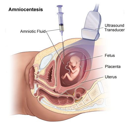
An amniocentesis is a procedure that involves inserting a long, thin needle through the mother's abdomen into the amniotic sac to withdraw a small sample of the amniotic fluid for examination. The amniotic fluid contains cells shed by the fetus, which contain genetic information. Although specific details of each procedure vary slightly, generally, an amniocentesis follows this process:
The woman's abdomen is cleansed with an antiseptic.
The doctor may or may not give a local anesthetic to numb the skin.
Ultrasound is used to help guide a hollow needle into the amniotic sac.
A small sample of fluid is withdrawn for laboratory analysis.
Strenuous activities should be avoided for 24 hours following an amniocentesis.
Women may feel some cramping during or after the amniocentesis.
Women with twins or other multiples need sampling from each amniotic sac, in order to study each baby. Depending on the position of the baby, placenta, amount of fluid, or mother's anatomy, sometimes the amniocentesis cannot be performed.
Women who are Rh negative will likely receive an Rh(D) immune globulin shot following amniocentesis.
The fluid is sent to a genetics laboratory so that the cells can grow and be analyzed. Alpha-fetoprotein, a protein made by the fetus that is present in the fluid, may also be measured to rule out an open neural tube defect, such as spina bifida. Results are usually available in about 10 days to two weeks, depending on the laboratory.
What are the risks and benefits of amniocentesis?
Risks |
Benefits |
|---|---|
After an amniocentesis, women may experience cramping, bleeding, or leaking of amniotic fluid. There is also a slight risk of infection. The risk of miscarriage is generally considered to be less than 1 percent after an amniocentesis in the second trimester of pregnancy. This is only slightly higher than the normal risk of miscarriage without an amniocentesis at this time in pregnancy. The American College of Obstetricians and Gynecologists does not recommend early amniocentesis (before 14 weeks), because the risks for miscarriage and pregnancy complications are higher than with amniocentesis performed between 15 and 17 weeks. However, some doctors may perform the procedure if a woman requests it. |
Amniocentesis helps confirm a tentative diagnosis of an abnormality found with other testing. It may also find that a fetus does not have the abnormality that was suspected. This allows couples to plan the remainder of pregnancy and to consider their options. Amniocentesis offers:
|

Connect with us:
Download our App: