nutch_noindex
CANCEL
COVID-2019 Alert

The latest information about the 2019 Novel Coronavirus, including vaccine clinics for children ages 6 months and older.

La información más reciente sobre el nuevo Coronavirus de 2019, incluidas las clínicas de vacunación para niños de 6 meses en adelante.

/nutch_noindex

Elizabeth Egan, MD

  • Elizabeth Sullivan Egan

Specialties

Infectious Diseases

Work and Education

Professional Education

Tufts University School of Medicine, Boston, MA, 5/22/2005

Internship

Boston Children's Hospital, Boston, MA, 6/30/2006

Residency

Boston Children's Hospital, Boston, MA, 6/30/2008

Fellowship

Boston Children's Hospital, Boston, MA, 6/30/2011

Board Certifications

Pediatric Infectious Disease
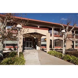
s, American Board of Pediatrics

Pediatrics, American Board of Pediatrics

All Publications

Erythrocyte-Plasmodium interactions: genetic manipulation of the erythroid lineage. Current opinion in microbiology Tetard, M., Peterson, N. A., Egan, E. S. 2022; 70: 102221

Abstract

Targeting critical host factors is an emerging concept in the treatment of infectious diseases. As obligate pathogens of erythrocytes, the Plasmodium spp. parasites that cause malaria must exploit erythroid host factors for their survival. However, our understanding of this important aspect of the malaria lifecycle is limited, in part because erythrocytes are enucleated cells that lack a nucleus and DNA, rendering them genetically intractable. Recent advances in genetic analysis of the erythroid lineage using small-hairpin RNAs and clustered regularly interspaced short palindromic repeats (CRISPR)/CRISPR-associated nuclease 9 (Cas9) in red-blood cells derived from stem cells have generated new insights into the functions of several candidate host factors for Plasmodium parasites. Along with efforts in other hematopoietic cells, these advances have also laid a strong foundation for genetic screens to identify novel erythrocyte host factors for malaria.

View details for DOI 10.1016/j.mib.2022.102221

View details for PubMedID 36242898

Cryo-electron tomography with mixed-scale dense neural networks reveals key steps in deployment of Toxoplasma invasion machinery. PNAS nexus Segev-Zarko, L. A., Dahlberg, P. D., Sun, S. Y., Pelt, D. M., Kim, C. Y., Egan, E. S., Sethian, J. A., Chiu, W., Boothroyd, J. C. 2022; 1 (4): pgac183

Abstract

Host cell invasion by intracellular, eukaryotic parasites within the phylum Apicomplexa is a remarkable and active process involving the coordinated action of apical organelles and other structures. To date, capturing how these structures interact during invasion has been difficult to observe in detail. Here, we used cryogenic electron tomography to image the apical complex of Toxoplasma gondii tachyzoites under conditions that mimic resting parasites and those primed to invade through stimulation with calcium ionophore. Through the application of mixed-scale dense networks for image processing, we developed a highly efficient pipeline for annotation of tomograms, enabling us to identify and extract densities of relevant subcellular organelles and accurately analyze features in 3-D. The results reveal a dramatic change in the shape of the anteriorly located apical vesicle upon its apparent fusion with a rhoptry that occurs only in the stimulated parasites. We also present information indicating that this vesicle originates from the vesicles that parallel the intraconoidal microtubules and that the latter two structures are linked by a novel tether. We show that a rosette structure previously proposed to be involved in rhoptry secretion is associated with apical vesicles beyond just the most anterior one. This result, suggesting multiple vesicles are primed to enable rhoptry secretion, may shed light on the mechanisms Toxoplasma employs to enable repeated invasion attempts. Using the same approach, we examine Plasmodium falciparum merozoites and show that they too possess an apical vesicle just beneath a rosette, demonstrating evolutionary conservation of this overall subcellular organization.

View details for DOI 10.1093/pnasnexus/pgac183

View details for PubMedID 36329726

View details for PubMedCentralID PMC9615128

Automated Recognition of Plasmodium falciparum Parasites from Portable Blood Levitation Imaging. Advanced science (Weinheim, Baden-Wurttemberg, Germany) Deshmukh, S. S., Byaruhanga, O., Tumwebaze, P., Akin, D., Greenhouse, B., Egan, E. S., Demirci, U. 2022: e2105396

Abstract

In many malaria-endemic regions, current detection tools are inadequate in diagnostic accuracy and accessibility. To meet the need for direct, phenotypic, and automated malaria parasite detection in field settings, a portable platform to process, image, and analyze whole blood to detect Plasmodium falciparum parasites, is developed. The liberated parasites from lysed red blood cells suspended in a magnetic field are accurately detected using this cellphone-interfaced, battery-operated imaging platform. A validation study is conducted at Ugandan clinics, processing 45malaria-negative and 36malaria-positive clinical samples without external infrastructure. Texture and morphology features are extracted from the sample images, and a random forest classifier is trained to assess infection status, achieving 100% sensitivity and 91% specificity against gold-standard measurements (microscopy and polymerase chain reaction), and limit of detection of 31parasites per L. This rapid and user-friendly platform enables portable parasite detection and can support malaria diagnostics, surveillance, and research in resource-constrained environments.

View details for DOI 10.1002/advs.202105396

View details for PubMedID 35957519

Revisiting the malaria hypothesis: accounting for polygenicity and pleiotropy. Trends in parasitology Ebel, E. R., Uricchio, L. H., Petrov, D. A., Egan, E. S. 1800

Abstract

The malaria hypothesis predicts local, balancing selection of deleterious alleles that confer strong protection from malaria. Three protective variants, recently discovered in red cell genes, are indeed more common in African than European populations. Still, up to 89% of the heritability of severe malaria is attributed to many genome-wide loci with individually small effects. Recent analyses of hundreds of genome-wide association studies (GWAS) in humans suggest that most functional, polygenic variation is pleiotropic for multiple traits. Interestingly, GWAS alleles and red cell traits associated with small reductions in malaria risk are not enriched in African populations. We propose that other selective and neutral forces, in addition to malaria prevalence, explain the global distribution of most genetic variation impacting malaria risk.

View details for DOI 10.1016/j.pt.2021.12.007

View details for PubMedID 35065882

Uncovering a Cryptic Site of Malaria Pathogenesis: Models to Study Interactions Between Plasmodium and the Bone Marrow. Frontiers in cellular and infection microbiology Feldman, T. P., Egan, E. S. 2022; 12: 917267

Abstract

The bone marrow is a critical site of host-pathogen interactions in malaria infection. The discovery of Plasmodium asexual and transmission stages in the bone marrow has renewed interest in the tissue as a niche for cellular development of both host and parasite. Despite its importance, bone marrow in malaria infection remains largely unexplored due to the challenge of modeling the complex hematopoietic environment in vitro. Advancements in modeling human erythropoiesis ex-vivo from primary human hematopoietic stem and progenitor cells provide a foothold to study the host-parasite interactions occurring in this understudied site of malaria pathogenesis. This review focuses on current in vitro methods to recapitulate and assess bone marrow erythropoiesis and their potential applications in the malaria field. We summarize recent studies that leveraged ex-vivo erythropoiesis to shed light on gametocyte development in nucleated erythroid stem cells and begin to characterize host cell responses to Plasmodium infection in the hematopoietic niche. Such models hold potential to elucidate mechanisms of disordered erythropoiesis, an underlying contributor to malaria anemia, as well as understand the biological determinants of parasite sexual conversion. This review compares the advantages and limitations of the ex-vivo erythropoiesis approach with those of in vivo human and animal studies of the hematopoietic niche in malaria infection. We highlight the need for studies that apply single cell analyses to this complex system and incorporate physical and cellular components of the bone marrow that may influence erythropoiesis and parasite development.

View details for DOI 10.3389/fcimb.2022.917267

View details for PubMedID 35719356

Common host variation drives malaria parasite fitness in healthy human red cells. eLife Ebel, E. R., Kuypers, F. A., Lin, C., Petrov, D. A., Egan, E. S. 2021; 10

Abstract

The replication of Plasmodium falciparum parasites within red blood cells (RBCs) causes severe disease in humans, especially in Africa. Deleterious alleles like hemoglobin S are well-known to confer strong resistance to malaria, but the effects of common RBC variation are largely undetermined. Here we collected fresh blood samples from 121 healthy donors, most with African ancestry, and performed exome sequencing, detailed RBC phenotyping, and parasite fitness assays. Over one third of healthy donors unknowingly carried alleles for G6PD deficiency or hemoglobinopathies, which were associated with characteristic RBC phenotypes. Among non-carriers alone, variation in RBC hydration, membrane deformability, and volume was strongly associated with P. falciparum growth rate. Common genetic variants in PIEZO1, SPTA1/SPTB, and several P. falciparum invasion receptors were also associated with parasite growth rate. Interestingly, we observed little or negative evidence for divergent selection on non-pathogenic RBC variation between Africans and Europeans. These findings suggest a model in which globally widespread variation in a moderate number of genes and phenotypes modulates P. falciparum fitness in RBCs.

View details for DOI 10.7554/eLife.69808

View details for PubMedID 34553687

Common host variation drives malaria parasite fitness in healthy human red cells ELIFE Ebel, E. R., Kuypers, F. A., Lin, C., Petrov, D. A., Egan, E. S. 2021; 10
Multiparametric biophysical profiling of red blood cells in malaria infection. Communications biology Deshmukh, S. S., Shakya, B., Chen, A., Durmus, N. G., Greenhouse, B., Egan, E. S., Demirci, U. 2021; 4 (1): 697

Abstract

Biophysical separation promises label-free, less-invasive methods to manipulate the diverse properties of live cells, such as density, magnetic susceptibility, and morphological characteristics. However, some cellular changes are so minute that they are undetectable by current methods. We developed a multiparametric cell-separation approach to profile cells with simultaneously changing density and magnetic susceptibility. We demonstrated this approach with the natural biophysical phenomenon of Plasmodium falciparum infection, which modifies its host erythrocyte by simultaneously decreasing density and increasing magnetic susceptibility. Current approaches have used these properties separately to isolate later-stage infected cells, but not in combination. We present biophysical separation of infected erythrocytes by balancing gravitational and magnetic forces to differentiate infected cell stages, including early stages for the first time, using magnetic levitation. We quantified height distributions of erythrocyte populations-27 ring-stage synchronized samples and 35 uninfected controls-and quantified their unique biophysical signatures. This platform can thus enable multidimensional biophysical measurements on unique cell types.

View details for DOI 10.1038/s42003-021-02181-3

View details for PubMedID 34103669

Erythrocyte CD55 mediates the internalization of Plasmodium falciparum parasites. eLife Shakya, B., Patel, S. D., Tani, Y., Egan, E. S. 2021; 10

Abstract

Invasion of human erythrocytes by the malaria parasite Plasmodium falciparum is a multi-step process. Previously, a forward genetic screen for P. falciparum host factors identified erythrocyte CD55 as essential for invasion, but its specific role and how it interfaces with the other factors that mediate this complex process are unknown. Using CRISPR-Cas9 editing, antibody-based inhibition, and live cell imaging, here we show that CD55 is specifically required for parasite internalization. Pre-invasion kinetics, erythrocyte deformability, and echinocytosis were not influenced by CD55, but entry was inhibited when CD55 was blocked or absent. Visualization of parasites attached to CD55-null erythrocytes point to a role for CD55 in stability and/or progression of the moving junction. Our findings demonstrate that CD55 acts after discharge of the parasite's rhoptry organelles, and plays a unique role relative to all other invasion receptors. As the requirement for CD55 is strain-transcendent, these results suggest that CD55 or its interacting partners may hold potential as therapeutic targets for malaria.

View details for DOI 10.7554/eLife.61516

View details for PubMedID 34028351

Mitochondria-Rich Extracellular Vesicles From Autologous Stem Cell-Derived Cardiomyocytes Restore Energetics of Ischemic Myocardium. Journal of the American College of Cardiology Ikeda, G. n., Santoso, M. R., Tada, Y. n., Li, A. M., Vaskova, E. n., Jung, J. H., O'Brien, C. n., Egan, E. n., Ye, J. n., Yang, P. C. 2021; 77 (8): 107388

Abstract

Mitochondrial dysfunction results in an imbalance between energy supply and demand in a failing heart. An innovative therapy that targets the intracellular bioenergetics directly through mitochondria transfer may be necessary.The purpose of this study was to establish a preclinical proof-of-concept that extracellular vesicle (EV)-mediated transfer of autologous mitochondria and their related energy source enhance cardiac function through restoration of myocardial bioenergetics.Human-induced pluripotent stem cell-derived cardiomyocytes (iCMs) were employed. iCM-conditioned medium was ultracentrifuged to collect mitochondria-rich EVs (M-EVs). Therapeutic effects of M-EVs were investigated using invivo murine myocardial infarction (MI) model.Electron microscopy revealed healthy-shaped mitochondria inside M-EVs. Confocal microscopy showed that M-EV-derived mitochondria were transferred into the recipient iCMs and fused with their endogenous mitochondrial networks. Treatment with 1.0 108/ml M-EVs significantly restored the intracellular adenosine triphosphate production and improved contractile profiles of hypoxia-injured iCMs as early as 3h after treatment. In contrast, isolated mitochondria that contained 300 more mitochondrial proteins than 1.0 108/ml M-EVs showed no effect after 24 h. M-EVs contained mitochondrial biogenesis-related messenger ribonucleic acids, including proliferator-activated receptor coactivator-1, which on transfer activated mitochondrial biogenesis in the recipient iCMs at 24h after treatment. Finally, intramyocardial injection of 1.0 108 M-EVs demonstrated significantly improved post-MI cardiac function through restoration of bioenergetics and mitochondrial biogenesis.M-EVs facilitated immediate transfer of their mitochondrial and nonmitochondrial cargos, contributing to improved intracellular energetics invitro. Intramyocardial injection of M-EVs enhanced post-MI cardiac function invivo. This therapy can be developed as a novel, precision therapeutic for mitochondria-related diseases including heart failure.

View details for DOI 10.1016/j.jacc.2020.12.060

View details for PubMedID 33632482

Mitochondria-Rich Extracellular Vesicles Rescue Patient-Specific Cardiomyocytes From Doxorubicin Injury: Insights Into the SENECA Trial. JACC. CardioOncology O'Brien, C. G., Ozen, M. O., Ikeda, G., Vaskova, E., Jung, J. H., Bayardo, N., Santoso, M. R., Shi, L., Wahlquist, C., Jiang, Z., Jung, Y., Zeng, Y., Egan, E., Sinclair, R., Gee, A., Witteles, R., Mercola, M., Svensson, K. J., Demirci, U., Yang, P. C. 2021; 3 (3): 428-440

Abstract

Anthracycline-induced cardiomyopathy (AIC) is a significant source of morbidity and mortality in cancer survivors. The role of mesenchymal stem cells (MSCs) in treating AIC was evaluated in the SENECA trial, a Phase 1 National Heart, Lung, and Blood Institute-sponsored study, but the mechanisms underpinning efficacy in human tissue need clarification.The purpose of this study was to perform an invitro clinical trial evaluating the efficacy and putative mechanisms of SENECA trial-specific MSCs in treating doxorubicin (DOX) injury, using patient-specific induced pluripotent stem cell-derived cardiomyocytes (iCMs) generated from SENECA patients.Patient-specific iCMs were injured with 1mol/L DOX for 24 hours, treated with extracellular vesicles (EVs) from MSCs by either coculture or direct incubation and then assessed for viability and markers of improved cellular physiology. MSC-derived EVs were separated into large extracellular vesicles (L-EVs) (>200nm) and small EVs (<220nm) using a novel filtration system.iCMs cocultured with MSCs in a transwell system demonstrated improved iCM viability and attenuated apoptosis. L-EVs but not small EVs recapitulated this therapeutic effect. L-EVs were found to be enriched in mitochondria, which were shown to be taken up by iCMs. iCMs treated with L-EVs demonstrated improved contractility, reactive oxygen species production, ATP production, and mitochondrial biogenesis. Inhibiting L-EV mitochondrial function with 1-methyl-4-phenylpyridinium attenuated efficacy.L-EV-mediated mitochondrial transfer mitigates DOX injury in patient-specific iCMs. Although SENECA was not designed to test MSC efficacy, consistent tendencies toward a positive effect were observed across endpoints. Our results suggest a mechanism by which MSCs may improve cardiovascular performance in AIC independent of regeneration, which could inform future trial design evaluating the therapeutic potential of MSCs.

View details for DOI 10.1016/j.jaccao.2021.05.006

View details for PubMedID 34604804

View details for PubMedCentralID PMC8463733

A common polymorphism in the mechanosensitive ion channel PIEZO1 is associated with protection from severe malaria in humans. Proceedings of the National Academy of Sciences of the United States of America Nguetse, C. N., Purington, N. n., Ebel, E. R., Shakya, B. n., Tetard, M. n., Kremsner, P. G., Velavan, T. P., Egan, E. S. 2020

Abstract

Malaria caused by the apicomplexan parasite Plasmodium falciparum has served as a strong evolutionary force throughout human history, selecting for red blood cell polymorphisms that confer innate protection against severe disease. Recently, gain-of-function mutations in the mechanosensitive ion channel PIEZO1 were shown to ameliorate Plasmodium parasite growth, blood-brain barrier dysfunction, and mortality in a mouse model of malaria. In humans, the gain-of-function allele PIEZO1 E756del is highly prevalent and enriched in Africans, raising the possibility that it is under positive selection due to malaria. Here we used a case-control study design to test for an association between PIEZO1 E756del and malaria severity among children in Gabon. We found that the E756del variant is strongly associated with protection against severe malaria in heterozygotes. In subjects with sickle cell trait, heterozygosity for PIEZO1 E756del did not confer additive protection and homozygosity was associated with an elevated risk of severe disease, suggesting an epistatic relationship between hemoglobin S and PIEZO1 E756del. Using donor blood samples, we show that red cells heterozygous for PIEZO1 E756del are not dehydrated and can support the intracellular growth of P. falciparum similar to wild-type cells. However, surface expression of the P. falciparum virulence protein PfEMP-1 was significantly reduced in infected cells heterozygous for PIEZO1 756del, a phenomenon that has been observed with other protective polymorphisms, such as hemoglobin C. Our findings demonstrate that PIEZO1 is an important innate determinant of malaria susceptibility in humans and suggest that the mechanism of protection may be related to impaired export of P. falciparum virulence proteins.

View details for DOI 10.1073/pnas.1919843117

View details for PubMedID 32265284

MICROSCALE MAGNETIC LEVITATION FOR MULTIPLEXED ANALYSIS OF MALARIA-INFECTED BLOOD SAMPLES IN RESOURCE-LIMITED SETTINGS Deshmukh, S. S., Durmus, N., Greenhouse, B., Egan, E., Demirci, U. AMER SOC TROP MED & HYGIENE. 2019: 13031
Beyond Hemoglobin: Screening for Malaria Host Factors TRENDS IN GENETICS Egan, E. S. 2018; 34 (2): 13341

Abstract

Severe malaria is caused by the Apicomplexan parasite Plasmodium falciparum, and results in significant global morbidity and mortality, particularly among young children and pregnant women. P. falciparum exclusively infects human erythrocytes during clinical illness, and several natural erythrocyte polymorphisms are protective against severe malaria. Since erythrocytes are enucleated and lack DNA, genetic approaches to understand erythrocyte determinants of malaria infection have historically been limited. This review highlights recent advances in the use of hematopoietic stem cells to facilitate genetic screening for malaria host factors. While challenges still exist, this approach holds promise for gaining new insights into host-pathogen interactions in malaria.

View details for PubMedID 29249333

Erythrocytes lacking the Langereis blood group protein ABCB6 are resistant to the malaria parasite Plasmodium falciparum. Communications biology Egan, E. S., Weekes, M. P., Kanjee, U., Manzo, J., Srinivasan, A., Lomas-Francis, C., Westhoff, C., Takahashi, J., Tanaka, M., Watanabe, S., Brugnara, C., Gygi, S. P., Tani, Y., Duraisingh, M. T. 2018; 1: 45

Abstract

The ATP-binding cassette transporter ABCB6 was recently discovered to encode the Langereis (Lan) blood group antigen. Lan null individuals are asymptomatic, and the function of ABCB6 in mature erythrocytes is not understood. Here, we assessed ABCB6 as a host factor for Plasmodium falciparum malaria parasites during erythrocyte invasion. We show that Lan null erythrocytes are highly resistant to invasion by P. falciparum, in a strain-transcendent manner. Although both Lan null and Jr(a-) erythrocytes harbor excess porphyrin, only Lan null erythrocytes exhibit a P. falciparum invasion defect. Further, the zoonotic parasite P. knowlesi invades Lan null and control cells with similar efficiency, suggesting that ABCB6 may mediate P. falciparum invasion through species-specific molecular interactions. Using tandem mass tag-based proteomics, we find that the only consistent difference in membrane proteins between Lan null and control cells is absence of ABCB6. Our results demonstrate that a newly identified naturally occurring blood group variant is associated with resistance to Plasmodium falciparum.

View details for PubMedID 30271928

CRISPR/Cas9 knockouts reveal genetic interaction between strain-transcendent erythrocyte determinants of &ITPlasmodium falciparum&IT invasion PROCEEDINGS OF THE NATIONAL ACADEMY OF SCIENCES OF THE UNITED STATES OF AMERICA Kanjee, U., Gruring, C., Chaand, M., Lin, K., Egan, E., Manzo, J., Jones, P. L., Yu, T., Barker, R., Weekes, M. P., Duraisingh, M. T. 2017; 114 (44): E9356E9365

Abstract

During malaria blood-stage infections, Plasmodium parasites interact with the RBC surface to enable invasion followed by intracellular proliferation. Critical factors involved in invasion have been identified using biochemical and genetic approaches including specific knockdowns of genes of interest from primary CD34+ hematopoietic stem cells (cRBCs). Here we report the development of a robust in vitro culture system to produce RBCs that allow the generation of gene knockouts via CRISPR/Cas9 using the immortal JK-1 erythroleukemia line. JK-1 cells spontaneously differentiate, generating cells at different stages of erythropoiesis, including terminally differentiated nucleated RBCs that we term "jkRBCs." A screen of small-molecule epigenetic regulators identified several bromodomain-specific inhibitors that promote differentiation and enable production of synchronous populations of jkRBCs. Global surface proteomic profiling revealed that jkRBCs express all known Pfalciparum host receptors in a similar fashion to cRBCs and that multiple Pfalciparum strains invade jkRBCs at comparable levels to cRBCs and RBCs. Using CRISPR/Cas9, we deleted two host factors, basigin (BSG) and CD44, for which no natural nulls exist. BSG interacts with the parasite ligand Rh5, a prominent vaccine candidate. A BSG knockout was completely refractory to parasite invasion in a strain-transcendent manner, confirming the essential role for BSG during invasion. CD44 was recently identified in an RNAi screen of blood group genes as a host factor for invasion, and we show that CD44 knockout results in strain-transcendent reduction in invasion. Furthermore, we demonstrate a functional interaction between these two determinants in mediating Pfalciparum erythrocyte invasion.

View details for DOI 10.1073/pnas.1711310114

View details for Web of Science ID 000414127400023

View details for PubMedID 29078358

View details for PubMedCentralID PMC5676921

Host-parasite interactions that guide red blood cell invasion by malaria parasites CURRENT OPINION IN HEMATOLOGY Paul, A. S., Egan, E. S., Duraisingh, M. T. 2015; 22 (3): 220-226

Abstract

Malaria is caused by the infection and proliferation of parasites from the genus Plasmodium in red blood cells (RBCs). A free Plasmodium parasite, or merozoite, released from an infected RBC must invade another RBC host cell to sustain a blood-stage infection. Here, we review recent advances on RBC invasion by Plasmodium merozoites, focusing on specific molecular interactions between host and parasite.Recent work highlights the central role of host-parasite interactions at virtually every stage of RBC invasion by merozoites. Biophysical experiments have for the first time measured the strength of merozoite-RBC attachment during invasion. For P. falciparum, there have been many key insights regarding the invasion ligand PfRh5 in particular, including its influence on host species tropism, a co-crystal structure with its RBC receptor basigin, and its suitability as a vaccine target. For P. vivax, researchers identified the origin and emergence of the parasite from Africa, demonstrating a natural link to the Duffy-negative RBC variant in African populations. For the simian parasite P. knowlesi, zoonotic invasion into human cells is linked to RBC age, which has implications for parasitemia during an infection and thus malaria.New studies of the molecular and cellular mechanisms governing RBC invasion by Plasmodium parasites have shed light on various aspects of parasite biology and host cell tropism, and indicate opportunities for malaria control.

View details for DOI 10.1097/MOH.0000000000000135

View details for Web of Science ID 000352793900005

View details for PubMedID 25767956

View details for PubMedCentralID PMC4418178

A forward genetic screen identifies erythrocyte CD55 as essential for Plasmodium falciparum invasion. SCIENCE Egan, E. S., Jiang, R. H., Moechtar, M. A., et al 2015; 348: 711-714

Abstract

Transmission of Plasmodium falciparum malaria parasites requires formation and development of gametocytes, yet all but the most mature of these sexual parasite forms are absent from the blood circulation. We performed a systematic organ survey in pediatric cases of fatal malaria to characterize the spatial dynamics of gametocyte development in the human host. Histological studies revealed a niche in the extravascular space of the human bone marrow where gametocytes formed in erythroid precursor cells and underwent development before reentering the circulation. Accumulation of gametocytes in the hematopoietic system of human bone marrow did not rely on cytoadherence to the vasculature as does sequestration of asexual-stage parasites. This suggests a different mechanism for the sequestration of gametocytes that could potentially be exploited to block malaria transmission.

View details for DOI 10.1126/scitranslmed.3008882

View details for Web of Science ID 000338713000005

View details for PubMedID 25009232

Plasmodium falciparum transmission stages accumulate in the human bone marrow SCIENCE TRANSLATIONAL MEDICINE Joice, R., Nilsson, S. K., Montgomery, J., Dankwa, S., Egan, E., Morahan, B., Seydel, K. B., Bertuccini, L., Alano, P., Williamson, K. C., Duraisingh, M. T., Taylor, T. E., Milner, D. A., Marti, M. 2014; 6 (244)

Abstract

Malaria remains a major cause of morbidity and mortality worldwide. Flow cytometry-based assays that take advantage of fluorescent protein (FP)-expressing malaria parasites have proven to be valuable tools for quantification and sorting of specific subpopulations of parasite-infected red blood cells. However, identification of rare subpopulations of parasites using green fluorescent protein (GFP) labelling is complicated by autofluorescence (AF) of red blood cells and low signal from transgenic parasites. It has been suggested that cell sorting yield could be improved by using filters that precisely match the emission spectrum of GFP.Detection of transgenic Plasmodium falciparum parasites expressing either tdTomato or GFP was performed using a flow cytometer with interchangeable optical filters. Parasitaemia was evaluated using different optical filters and, after optimization of optics, the GFP-expressing parasites were sorted and analysed by microscopy after cytospin preparation and by imaging cytometry.A new approach to evaluate filter performance in flow cytometry using two-dimensional dot blot was developed. By selecting optical filters with narrow bandpass (BP) and maximum position of filter emission close to GFP maximum emission in the FL1 channel (510/20, 512/20 and 517/20; dichroics 502LP and 466LP), AF was markedly decreased and signal-background improve dramatically. Sorting of GFP-expressing parasite populations in infected red blood cells at 90 or 95% purity with these filters resulted in 50-150% increased yield when compared to the standard filter set-up. The purity of the sorted population was confirmed using imaging cytometry and microscopy of cytospin preparations of sorted red blood cells infected with transgenic malaria parasites.Filter optimization is particularly important for applications where the FP signal and percentage of positive events are relatively low, such as analysis of parasite-infected samples with in the intention of gene-expression profiling and analysis. The approach outlined here results in substantially improved yield of GFP-expressing parasites, and requires decreased sorting time in comparison to standard methods. It is anticipated that this protocol will be useful for a wide range of applications involving rare events.

View details for DOI 10.1186/1475-2875-11-312

View details for Web of Science ID 000313865200001

View details for PubMedID 22950515

Optimization of flow cytometric detection and cell sorting of transgenic Plasmodium parasites using interchangeable optical filters MALARIA JOURNAL Vorobjev, I. A., Buchholz, K., Prabhat, P., Ketman, K., Egan, E. S., Marti, M., Duraisingh, M. T., Barteneva, N. S. 2012; 11

Abstract

Although the two Vibrio cholerae chromosomes initiate replication in a coordinated fashion, we show here that each chromosome appears to have a specific replication initiator. DnaA overproduction promoted overinitiation of chromosome I and not chromosome II. In contrast, overproduction of RctB, a protein that binds to the origin of replication of chromosome II, promoted overinitiation of chromosome II and not chromosome I.

View details for DOI 10.1128/JB.00565-06

View details for Web of Science ID 000240250200043

View details for PubMedID 16923911

View details for PubMedCentralID PMC1595377

Independent control of replication initiation of the two Vibrio cholerae chromosomes by DnaA and RctB JOURNAL OF BACTERIOLOGY Duigou, S., Knudsen, K. G., Skovgaard, O., Egan, E. S., Lobner-Olesen, A., Waldor, M. K. 2006; 188 (17): 6419-6424

Abstract

The RctB protein binds to the origin of replication of Vibrio cholerae chromosome II (chrII) and is required for oriCIIVc-based replication. Here, we found that RctB acts as an autorepressor, inhibiting rctB transcription. Integration host factor promotes rctB transcription, while Dam and DnaA, factors required for replication of both V. cholerae chromosomes, influence RctB autorepression. Thus, RctB appears to regulate chrII replication as both an initiator and a transcription repressor, and its synthesis is modulated by factors that govern replication of both chromosomes.

View details for Web of Science ID 000234677400045

View details for PubMedID 16385068

Autorepression of RctB, an initiator of Vibrio cholerae chromosome II replication JOURNAL OF BACTERIOLOGY Egan, E. S., Duigou, S., Waldor, M. K. 2006; 188 (2): 789-793

Abstract

Historically, the prokaryotic genome was assumed to consist of a single circular replicon. However, as more microbial genome sequencing projects are completed, it is becoming clear that multipartite genomes comprised of more than one chromosome are not unusual among prokaryotes. Chromosomes are distinguished from plasmids by the presence of essential genes as well as characteristic cell cycle-linked replication kinetics; unlike plasmids, chromosomes initiate replication once per cell cycle. The existence of multipartite prokaryotic genomes raises several questions regarding how multiple chromosomes are replicated and segregated during the cell cycle. These divided genomes also introduce questions regarding chromosome evolution and genome stability. In this review, we discuss these and other issues, with particular emphasis on the cholera pathogen Vibrio cholerae.

View details for DOI 10.1111/j.1365-2958.2005.04622.x

View details for Web of Science ID 000228975300002

View details for PubMedID 15882408

Divided genomes: negotiating the cell cycle in prokaryotes with multiple chromosomes MOLECULAR MICROBIOLOGY Egan, E. S., Fogel, M. A., Waldor, M. K. 2005; 56 (5): 1129-1138

View details for Web of Science ID 000222673700007

View details for PubMedID 15242627

Synchronous replication initiation of the two Vibrio cholerae chromosomes CURRENT BIOLOGY Egan, E. S., Lobner-Olesen, A., Waldor, M. K. 2004; 14 (13): R501-R502

Abstract

Studies of prokaryotic chromosome replication have focused almost exclusively on organisms with one chromosome. We defined and characterized the origins of replication of the two Vibrio cholerae chromosomes, oriCI(vc) and oriCII(vc). OriCII(vc) differs from the origin assigned by bioinformatic analysis and is unrelated to oriCI(vc). OriCII(vc)-based replication requires an internal 12 base pair repeat and two hypothetical genes that flank oriCII(vc). One of these genes is conserved among diverse genera of the family Vibrionaceae and encodes an origin binding protein. The other gene codes for an RNA and not a protein. OriCII(vc)- but not oriCI(vc)-based replication is negatively regulated by a DNA sequence adjacent to oriCII(vc). There is an unprecedented requirement for DNA adenine methyltransferase in both oriCI(vc)- and oriCII(vc)-based replication. Our studies of replication in V. cholerae indicate that microorganisms having multiple chromosomes may utilize unique mechanisms for the control of replication.

View details for Web of Science ID 000184928800014

View details for PubMedID 12941279

Distinct replication requirements for the two vibrio cholerae chromosomes CELL Egan, E. S., Waldor, M. K. 2003; 114 (4): 521-530

Abstract

Photic entrainment of insect circadian rhythms can occur through either extraretinal (brain) or retinal photoreceptors, which mediate sensitivity to blue light or longer wavelengths, respectively. Although visual transduction processes are well understood in the insect retina, almost nothing is known about the extraretinal blue light photoreceptor of insects. We now have identified and characterized a candidate blue light photoreceptor gene in Drosophila (DCry) that is homologous to the cryptochrome (Cry) genes of mammals and plants. The DCry gene is located in region 91F of the third chromosome, an interval that does not contain other genes required for circadian rhythmicity. The protein encoded by DCry is approximately 50% identical to the CRY1 and CRY2 proteins recently discovered in mammalian species. As expected for an extraretinal photoreceptor mediating circadian entrainment, DCry mRNA is expressed within the adult brain and can be detected within body tissues. Indeed, tissue in situ hybridization demonstrates prominent expression in cells of the lateral brain, which are close to or coincident with the Drosophila clock neurons. Interestingly, DCry mRNA abundance oscillates in a circadian manner in Drosophila head RNA extracts, and the temporal phasing of the rhythm is similar to that documented for the mouse Cry1 mRNA, which is expressed in clock tissues. Finally, we show that changes in DCry gene dosage are associated predictably with alterations of the blue light resetting response for the circadian rhythm of adult locomotor activity.

View details for Web of Science ID 000080162400003

View details for PubMedID 10233998

An extraretinally expressed insect cryptochrome with similarity to the blue light photoreceptors of mammals and plants JOURNAL OF NEUROSCIENCE Egan, E. S., Franklin, T. M., Hilderbrand-Chae, M. J., McNeil, G. P., Roberts, M. A., Schroeder, A. J., Zhang, X. L., Jackson, F. R. 1999; 19 (10): 3665-3673

Abstract

Genetic screens in zebrafish (Danio rerio) have isolated mutations in hundreds of genes with essential functions. To facilitate the identification of candidate genes for these mutations, we have genetically mapped 104 genes and expressed sequence tags by scoring single-strand conformational polymorphisms in a panel of haploid siblings. To integrate this map with existing genetic maps, we also scored 275 previously mapped genes, microsatellites, and sequence-tagged sites in the same haploid panel. Systematic phylogenetic analysis defined likely mammalian orthologs of mapped zebrafish genes, and comparison of map positions in zebrafish and mammals identified significant conservation of synteny. This comparative analysis also identified pairs of zebrafish genes that appear to be orthologous to single mammalian genes, suggesting that these genes arose in a genome duplication that occurred in the teleost lineage after the divergence of fish and mammal ancestors. This comparative map analysis will be useful in predicting the locations of zebrafish genes from mammalian gene maps and in understanding the evolution of the vertebrate genome.

View details for Web of Science ID 000079904300003

View details for PubMedID 10207156

A genetic linkage map for zebrafish: Comparative analysis and localization of genes and expressed sequences GENOME RESEARCH GATES, M. A., Kim, L., Egan, E. S., Cardozo, T., Sirotkin, H. I., Dougan, S. T., Lashkari, D., Abagyan, R., Schier, A. F., Talbot, W. S. 1999; 9 (4): 334-347

Abstract

The vertebrate body plan is established during gastrulation, when cells move inwards to form the mesodermal and endodermal germ layers. Signals from a region of dorsal mesoderm, which is termed the organizer, pattern the body axis by specifying the fates of neighbouring cells. The organizer is itself induced by earlier signals. Although members of the transforming growth factor-beta (TGF-beta) and Wnt families have been implicated in the formation of the organizer, no endogenous signalling molecule is known to be required for this process. Here we report that the zebrafish squint (sqt) and cyclops (cyc) genes have essential, although partly redundant, functions in organizer development and also in the formation of mesoderm and endoderm. We show that the sqt gene encodes a member of the TGF-beta superfamily that is related to mouse nodal. cyc encodes another nodal-related proteins, which is consistent with our genetic evidence that sqt and cyc have overlapping functions. The sqt gene is expressed in a dorsal region of the blastula that includes the extraembryonic yolk syncytial layer (YSL). The YSL has been implicated as a source of signals that induce organizer development and mesendoderm formation. Misexpression of sqt RNA within the embryo or specifically in the YSL induces expanded or ectopic dorsal mesoderm. These results establish an essential role for nodal-related signals in organizer development and mesendoderm formation.

View details for Web of Science ID 000075829900044

View details for PubMedID 9744277

Zebrafish organizer development and germ-layer formation require nodal-related signals NATURE Feldman, B., GATES, M. A., Egan, E. S., Dougan, S. T., Rennebeck, G., Sirotkin, H. I., Schier, A. F., Talbot, W. S. 1998; 395 (6698): 181-185

Abstract

Genes essential for vertebrate body plan specification, organ development, and organ function are likely to be shared between mammals and zebrafish, but only in zebrafish have large-scale, genome-wide mutagenesis screens been conducted to isolate embryonic lethal mutations. Discovering the roles played by these disrupted genes requires their molecular characterization, which would be facilitated by assaying large cloned genomic DNAs for their potential to rescue mutant phenotypes. Here we demonstrate that bacterial artificial chromosomes can rescue the phenotype of floating head (flh) mutants. Homozygous flh embryos lack a differentiated notochord and have a reduced, discontinuous floor plate. Mutant embryos injected with genomic clones containing the flh+ gene often had stretches of several to many notochord cells overlaid by a row of floor-plate cells. In contrast, control mutant embryos injected with artificial chromosomes lacking the flh+ gene failed to form notochord. We conclude that the injection of large-insert genomic clones will speed the isolation of zebrafish genes disrupted by mutation and hence the identification of gene functions necessary for development of vertebrate embryos.

View details for Web of Science ID 000074367700018

View details for PubMedID 9653657

Mutant rescue by BAC clone injection in zebrafish GENOMICS Yan, Y. L., Talbot, W. S., Egan, E. S., Postlethwait, J. H. 1998; 50 (2): 287-289

Abstract

In chordate phylogeny, changes in the nervous system, jaws, and appendages transformed meek filter feeders into fearsome predators. Gene duplication is thought to promote such innovation. Vertebrate ancestors probably had single copies of genes now found in multiple copies in vertebrates and gene maps suggest that this occurred by polyploidization. It has been suggested that one genome duplication event occurred before, and one after the divergence of ray-finned and lobe-finned fishes. Holland et al., however, have argued that because various vertebrates have several HOX clusters, two rounds of duplication occurred before the origin of jawed fishes. Such gene-number data, however, do not distinguish between tandem duplications and polyploidization events, nor whether independent duplications occurred in different lineages. To investigate these matters, we mapped 144 zebrafish genes and compared the resulting map with mammalian maps. Comparison revealed large conserved chromosome segments. Because duplicated chromosome segments in zebrafish often correspond with specific chromosome segments in mammals, it is likely that two polyploidization events occurred prior to the divergence of fish and mammal lineages. This zebrafish gene map will facilitate molecular identification of mutated zebrafish genes, which can suggest functions for human genes known only by sequence.

View details for Web of Science ID 000072755500016

View details for PubMedID 9537416

Vertebrate genome evolution and the zebrafish gene map NATURE GENETICS Postlethwait, J. H., Yan, Y. L., GATES, M. A., Horne, S., Amores, A., Brownlie, A., Donovan, A., Egan, E. S., Force, A., Gong, Z. Y., Goutel, C., Fritz, A., Kelsh, R., Knapik, E., Liao, E., Paw, B., Ransom, D., Singer, A., Thomson, M., Abduljabbar, T. S., Yelick, P., Beier, D., Joly, J. S., Larhammar, D., ROSA, F., Westerfield, M., Zon, L. I., Johnson, S. L., Talbot, W. S. 1998; 18 (4): 345-349

Abstract

Genetic screens in zebrafish have provided mutations in hundreds of genes with essential functions in the developing embryo. To investigate the possible uses of chromosomal rearrangements in the analysis of these mutations, we genetically characterized three gamma-ray induced alleles of cyclops (cyc), a gene required for development of midline structures. We show that cyc maps near one end of Linkage Group 12 (LG 12) and that this region is involved in a reciprocal translocation with LG 2 in one gamma-ray induced mutation, cyc(b213). The translocated segments together cover approximately 5% of the genetic map, and we show that this rearrangement is useful for mapping cloned genes that reside in the affected chromosomal regions. The other two alleles, cyc(b16) and cyc(b229), have deletions in the distal region of LG 12. Interestingly, both of these mutations suppress recombination between genetic markers in LG 12, including markers at a distance from the deletion. This observation raises the possibility that these deletions affect a site required for meiotic recombination on the LG 12 chromosome. The cyc(b16) and cyc(b229) mutations may be useful for balancing other lethal mutations located in the distal region of LG 12. These results show that chromosomal rearrangements can provide useful resources for mapping and genetic analyses in zebrafish.

View details for Web of Science ID 000071494000034

View details for PubMedID 9475747

View details for PubMedCentralID PMC1459804

Genetic analysis of chromosomal rearrangements in the cyclops region of the zebrafish genome GENETICS Talbot, W. S., Egan, E. S., GATES, M. A., Walker, C., Ullmann, B., Neuhauss, S. C., Kimmel, C. B., Postlethwait, J. H. 1998; 148 (1): 373-380