Notice of West building lobby closure at Lucile Packard Children’s Hospital Stanford

Gordon Bae, MD
Clinical Assistant Professor
Dermatology
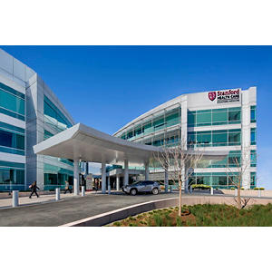
Stanford Medicine Outpatient Center
Stanford Dermatology
450 Broadway Street
Redwood City, CA 94063
Phone:
(844) 416-7846
Fax:
(650) 721-3476
Locations
Stanford Dermatology
450 Broadway Street
Redwood City, CA 94063
Phone : (844) 416-7846
Fax : (650) 721-3476
Work and Education
Professional Education
Harvard Medical School, Boston, MA, 6/1/2016
Residency
Stanford University Dermatology Residency, Redwood City, CA, 6/30/2020
Internship
Brigham and Women's Hospital Internal Medicine Residency, Boston, MA, 6/30/2017
Board Certifications
Dermatology, American Board of Dermatology, 2020
Languages
English
Korean
Spanish

Connect with us:
Download our App: