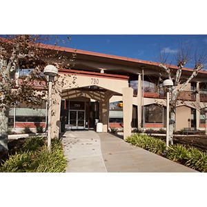
1st Fl
Palo Alto, CA 94304
Fax: (650) 497-8339
Working with children and their families is an honor and a privilege. Children are truly magical in how they soak in the world. I love being part of a community that helps to keep them healthy and happy. As a parent, I know how important it is to build a relationship on trust with your doctors. Its very important for me to learn about each patient and their family so that we can take the best care of your child together. I love that at Stanford Children's we are always discovering innovative and personalized care centered around your child to ensure that they can continue to grow and explore the world.
As a pediatric otolaryngologist, I specialize in diseases of the head and neck in children, including ear infections, hearing loss, sinus issues, sleep apnea, airway concerns, and voice/swallow problems. My goal is to educate and empower children and their families so that together we can make the best decision for your child.
Stanford University School of Medicine, Palo Alto, CA, 06/01/2015
Stanford University Otolaryngology Residency, Stanford, CA, 06/30/2016
Stanford University Otolaryngology Residency, Stanford, CA, 06/30/2022
Stanford University Pediatric Otolaryngology Fellowship, Stanford, CA, 06/30/2023
SignificanceThe mammalian utricle shows limited hair cell regeneration despite partial recovery of function. Recovery of vestibular evoked potentials, a measure of utricular function, occurs after utricular hair cell damage in mammals. While previous work has shown limited regeneration in response to damage, most regenerating hair cells are type II with immature-appearing bundles. Whether hair cells that remain ("surviving hair cells") can self-repair and contribute to functional recovery is unknown. Using lineage tracing, we have characterized surviving hair cells over time and found that they repair bundles, regain innervation, and remain differentiated, and are therefore poised to contribute to the recovery of organ function.
View details for DOI 10.1073/pnas.2116973119
View details for PubMedID 35380897
OBJECTIVES: To review the outcomes of repairing tegmen dehiscence using the middle cranial fossa approach with a self-setting bone cement.STUDY DESIGN: Retrospective case series.SETTING: Two academic tertiary hospitals.PATIENTS: All patients presenting for surgical repair of tegmen dehiscence and with postoperative follow-up for at least 6months between October 2015 and July 2019.INTERVENTION: Surgical repair using a middle cranial fossa approach using a layered reconstruction with temporalis fascia and self-setting calcium phosphate bone cement.MAIN OUTCOME MEASURES: Perioperative complications, recurrence of presenting symptoms/disease, hearing, and facial nerve grade.RESULTS: The cohort consisted of 22 patients with 23 tegmen dehiscence repairs (1 sequential bilateral repair). There were 16 males and 6 females with an average age at operation of 52.6years. Repairs were left sided in 9, right sided in 12 patients, and bilateral in 1 patient. No patients had recurrence of presenting symptoms or disease at most recent follow-up. Preoperative hearing was maintained in all patients. Two patients (9% of repairs) experienced delayed partial temporary facial nerve weakness House-Brackman grade 2 and 4 which had recovered by 8weeks postoperative.CONCLUSION: We demonstrate a technique for repairing tegmen dehiscence of the middle cranial fossa floor that has excellent postoperative outcomes. We highlight potential technical challenges in this approach as well as the need for counseling for potential partial transient facial nerve dysfunction.
View details for DOI 10.1097/MAO.0000000000003110
View details for PubMedID 33710151
INTRODUCTION: The diagnosis of ankyloglossia, or tongue-tie, and the number of frenotomies performed has increased over 10-fold from 1997 to 2012 in the United States. The sharpest increase has been in neonates. For parents considering frenotomy for their breastfeeding newborn, there is controversy surrounding the evaluation of tongue-tie and the benefit of a frenotomy. Complications from tongue-tie procedures are thought to be low, though it is not well reported nor studied.OBJECTIVES: The aim of this study is to describe a case of a sublingual mucocele after laser frenotomy in a neonate with tongue-tie and to investigate major complications reported after tongue-tie release in pediatric patients through a systematic review of the literature.CASE REPORT: We present a 6-week-old female who underwent a laser frenotomy procedure performed by a dentist who presented with a new cyst under her tongue.MATERIAL AND METHODS: A systematic literature search of articles published from 1965 to April 2020 was conducted in Ovid MEDLINE(R), Ovid EMBASE, and Scopus. Citations were uploaded into a systematic review software program (DistillerSR, Ottawa, ON, Canada), followed by full text screening.RESULTS: 47 major complications were reported in 34 patients, including our patient. Most of the cases were located in the United States and Europe. The most frequent indications for the procedure were breastfeeding problems (n=18) and speech impediment (n=4). The procedure was performed by dentists (n=6), lactation consultants (n=5), and otolaryngologists (n=4). The bulk of the major complications after frenotomy included poor feeding (n=7), hypovolemic shock (n=4), apnea (n=4), acute airway obstruction (n=4), and Ludwig angina (n=2).CONCLUSIONS: Reporting of complications after frenotomy is lacking. Risks to neonates may be different than risks to older children and adults. Practitioners across different specialties should be monitoring and studying this more rigorously to better guide patients and families on the risks and benefits of this procedure.
View details for DOI 10.1016/j.ijporl.2020.110356
View details for PubMedID 32927351
View details for DOI 10.1007/s40136-020-00300-y
View details for Web of Science ID 000702480100011
View details for DOI 10.1002/lary.28921
View details for PubMedID 33059385
To estimate the prevalence and significance of cranial nerve (CN) imaging abnormalities in patients with hereditary neuropathy and discuss clinical implications.We retrospectively analyzed data from patients at four tertiary academic medical centers with hereditary neuropathy diagnoses who had undergone gadolinium-enhanced magnetic resonance imaging (MRI) of the brain or skull base between 2004 and 2018. MRI scans, as well as computed tomography imaging when available, were reviewed and bivariable analysis was performed to identify predictors of CN abnormalities on imaging.Among 39 patients meeting study criteria, 11 had clinical CN deficits (28%) and 8 had CN abnormalities on imaging (21%). Of the patients with CN abnormalities on imaging, half had CN deficits (4/8) and only a quarter had imaging abnormalities of the CNs with the deficits (2/8). Imaging abnormalities were found in varied CNs, including CNs III, V, VII, and the VII/VIII complex in the internal auditory canal. MRI obtained for the purpose of evaluating CN deficits had a statistically significant increased likelihood of containing CN imaging abnormalities. However, CN deficits themselves were not predictive of imaging abnormalities.Thickening and enhancement of CNs on MRI may be found in approximately 1/5 of patients with hereditary neuropathies and are inconsistently associated with clinical deficits. These imaging findings should not be mistaken for neoplastic and infectious processes as they may be manifestations of the patients' underlying genetic neuropathy.4.
View details for DOI 10.1002/lio2.343
View details for PubMedID 32128425
View details for PubMedCentralID PMC7042653
INTRODUCTION: Sound is integral to communication and connects us to the world through speech and music. Cochlear hair cells are essential for converting sounds into neural impulses. However, these cells are highly susceptible to damage from an array of factors, resulting in degeneration and ultimately irreversible hearing loss in humans. Since the discovery of hair cell regeneration in birds, there have been tremendous efforts to identify therapies that could promote hair cell regeneration in mammals. Areas covered: Here, we will review recent studies describing spontaneous hair cell regeneration and direct cellular reprograming as well as other factors that mediate mammalian hair cell regeneration. Expert opinion: Numerous combinatorial approaches have successfully reprogrammed non-sensory supporting cells to form hair cells, albeit with limited efficacy and maturation. Studies on epigenetic regulation and transcriptional network of hair cell progenitors may accelerate discovery of more promising reprogramming regimens.
View details for PubMedID 30584811
PURPOSE OF REVIEW: The primary purpose of this review is to summarize current literature in the field of vestibular regeneration with a focus on recent developments in molecular and gene therapies.RECENT FINDINGS: Since the discovery of limited vestibular hair cell regeneration in mammals in the 1990s, many elegant studies have improved our knowledge of mechanisms of development and regeneration of the vestibular system. A better understanding of the developmental pathways of the vestibular organs has fueled various biological strategies to enhance regeneration, including novel techniques in deriving vestibular hair cells from embryonic and induced pluripotent stem cells. In addition, the identification of specific genetic mutations responsible for vestibular disorders has opened various opportunities for gene replacement therapy.SUMMARY: Vestibular dysfunction is a significant clinical problem with limited therapeutic options, warranting research on biological strategies to repair/regenerate the vestibular organs to restore function. The use of gene therapy appears promising in animal models of vestibular dysfunction.
View details for DOI 10.1097/MOO.0000000000000477
View details for PubMedID 30045104
In medically refractory chronic frontal sinusitis, ethmoidectomy without instrumentation of the frontal ostium may resolve frontal disease. Our aim was to determine the efficacy of ethmoidectomy alone for the treatment of chronic frontal sinusitis.Adults with chronic rhinosinusitis prospectively enrolled in a multicenter study who demonstrated frontal sinusitis on computed tomography were divided into 2 groups: (1) endoscopic sinus surgery (ESS) incorporating ethmoidectomy, but excluding frontal sinusotomy; and (2) ESS incorporating frontal sinusotomy. The primary outcome was improvement in 22-item Sino-Nasal Outcome Test (SNOT-22) scores. Secondary outcomes included endoscopic scores and use of corticosteroids and antibiotics.A total of 196 cases undergoing frontal sinusotomy and 30 cases treated with ethmoidectomy without frontal sinusotomy were analyzed and were comparable demographically. The prevalence of nasal polyps, previous ESS, asthma, and aspirin intolerance was more common in the frontal sinusotomy group (p < 0.050). Preoperative endoscopy and computed tomography scores were higher in the frontal sinusotomy group (p 0.001). Postoperatively, both groups showed comparable SNOT-22 scores with worse endoscopy scores in the frontal sinusotomy group (p = 0.038). Postoperative improvement in SNOT-22 total and subdomain scores was comparable between groups. Nasal endoscopy scores improved to a greater degree in the frontal sinusotomy group (p = 0.023). Duration of postoperative topical steroid use was higher in the frontal sinusotomy group (p = 0.007). Revision surgery was needed in 2.6% of frontal sinusotomy patients and 0% of patients without frontal sinusotomy.The treatment of chronic frontal sinusitis through ethmoidectomy is a potential alternative to frontal sinusotomy achieving similar quality of life (QOL) improvements in patients manifesting less severe sinus disease.
View details for DOI 10.1002/alr.21726
View details for PubMedID 26879467
Recruitment of endogenous progenitors is critical during tissue repair. The inner ear utricle requires mechanosensory hair cells (HCs) to detect linear acceleration. After damage, non-mammalian utricles regenerate HCs via both proliferation and direct transdifferentiation. In adult mammals, limited transdifferentiation from unidentified progenitors occurs to regenerate extrastriolar Type II HCs. Here we show that HC damage in neonatal mouse utricle activates the Wnt target gene Lgr5 in striolar supporting cells. Lineage tracing and time-lapse microscopy reveal that Lgr5+ cells transdifferentiate into HC-like cells in vitro. In contrast to adults, HC ablation in neonatal utricles in vivo recruits Lgr5+ cells to regenerate striolar HCs through mitotic and transdifferentiation pathways. Both Type I and II HCs are regenerated, and regenerated HCs display stereocilia and synapses. Lastly, stabilized -catenin in Lgr5+ cells enhances mitotic activity and HC regeneration. Thus Lgr5 marks Wnt-regulated, damage-activated HC progenitors and may help uncover factors driving mammalian HC regeneration.
View details for DOI 10.1038/ncomms7613
View details for PubMedID 25849379
Wnt signaling is a highly conserved pathway crucial for development and homeostasis of multicellular organisms. Secreted Wnt ligands bind Frizzled receptors to regulate diverse processes such as axis patterning, cell division, and cell fate specification. They also serve to govern self-renewal of somatic stem cells in several adult tissues. The complexity of the pathway can be attributed to the myriad of Wnt and Frizzled combinations as well as its diverse context-dependent functions. In the developing mouse inner ear, Wnt signaling plays diverse roles, including specification of the otic placode and patterning of the otic vesicle. At later stages, its activity governs sensory hair cell specification, cell cycle regulation, and hair cell orientation. In regenerating sensory organs from non-mammalian species, Wnt signaling can also regulate the extent of proliferative hair cell regeneration. This review describes the current knowledge of the roles of Wnt signaling and Wnt-responsive cells in hair cell development and regeneration. We also discuss possible future directions and the potential application and limitation of Wnt signaling in augmenting hair cell regeneration.
View details for DOI 10.3389/fncel.2015.00066
View details for PubMedID 25814927