Notice of West building lobby closure at Lucile Packard Children’s Hospital Stanford
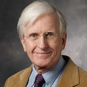
Michael Gaynon, MD
Clinical Professor
Ophthalmology
Watson Court – Palo Alto
Byers Eye Institute at Stanford
2452 Watson Court, Suite 1500
Palo Alto, CA 94303
Phone:
(650) 723-6995
Locations
Byers Eye Institute at Stanford
2452 Watson Court, Suite 1500
Palo Alto, CA 94303
Phone : (650) 723-6995
Work and Education
Professional Education
University of North Carolina at Chapel Hill, Chapel Hill, NC, 5/31/1972
Residency
Alton Ochsner Med Foundation, New Orleans, LA, 6/30/1976
Fellowship
Massachusetts Eye and Ear Infirmary Ophthalmic Training, Boston, MA, 12/31/1978
Internship
California Pacific Medical Center Internal Medicine Residency, San Francisco, CA, 6/30/1973
Board Certifications
Ophthalmology, American Board of Ophthalmology, 1978
Languages
English

Connect with us:
Download our App: