Notice of West building lobby closure at Lucile Packard Children’s Hospital Stanford
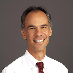
Nikolas Blevins, MD
Director, Stanford/LPCH Cochlear Implant Program | Professor
Neurotology
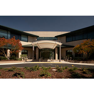
Stanford Ear Institute
2452 Watson Court, Suite 1500
Palo Alto, CA 94303
Phone:
(650) 498-4327
Fax:
(650) 736-4327
Locations
Work and Education
Professional Education
Harvard Medical School, Boston, MA, 06/30/1988
Residency
UCSF Dept of Otolaryngology Head and Neck Surgery, San Francisco, CA, 06/30/1994
Fellowship
UCSF Dept of Otolaryngology Head and Neck Surgery, San Francisco, CA, 06/30/1995
Internship
UCSF Dept of General Surgery, San Francisco, CA, 06/30/1990
Board Certifications
Otolaryngology, American Board of Otolaryngology, 1995
Neurotology, American Board of Otolaryngology, 2006
Languages
English

Connect with us:
Download our App: