Notice of West building lobby closure at Lucile Packard Children’s Hospital Stanford
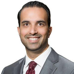
Vivek Buch, MD
Assistant Professor
Neurosurgery
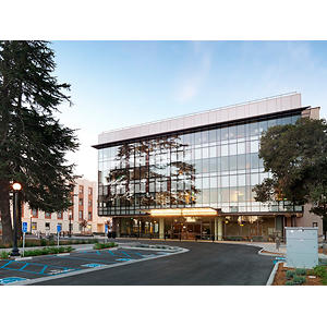
Stanford Neuroscience Health Center
213 Quarry Road, 1st Floor
Palo Alto, CA 94304
Phone:
(650) 725-5792
Fax:
(650) 725-5032
Locations
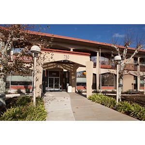
Pediatric Neurosurgery
730 Welch Road, 2nd floor
Palo Alto, CA 94304
Phone : (650) 723-0991
Fax : (650) 725-5086
Conditions
Epilepsy
Epilepsy Surgery
Work and Education
Professional Education
Warren Alpert Medical School Brown University, Providence, RI, 05/26/2013
Residency
University of Pennsylvania, Philadelphia, PA, 06/30/2020
Fellowship
Stanford University Dept of Neurosurgery, Palo Alto, CA, 06/30/2021
University of Pennsylvania Dept of Neurology, Philadelphia, PA, 12/31/2018
Languages
English
Gujarati

Connect with us:
Download our App: