nutch_noindex
Search Icon
    Menu Button
    Stanford Children's Health
    DonateContactMyChart LoginFind a Doctor
    • Second Opinion
    • Donate
    • Contact
    • Refer a Patient
    • En Español
    • Our Locations
    • Medical Services
    • About Us
    • Research & Innovation
    • Patients & Visitors
    • For Health Professionals
    • Second Opinion
    • Donate
    • En Español
    • MyChart Login
    • Recently Visited
      • Our Locations
      • Medical Services
      • About Us
      • Research & Innovation
      • Patients & Visitors
      • For Health Professionals
      CANCEL
      • View More Results

      Loading...

      COVID-2019 Alert

      Information about the 2019 Novel Coronavirus. Read the latest >

      Información sobre el coronavirus 2019 (COVID-19). Aprenda más >

      /nutch_noindex


      Anatomy of the Bladder

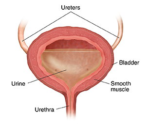
      The bladder is part of your urinary tract. It's a hollow organ in your lower belly (pelvis). Urine is stored in it. This is the liquid waste that’s made by the kidneys. Urine flows away from each kidney through a tube called a ureter. The ureters carry the urine into your bladder. The urine stays in your bladder until you let it pass out of your body through another tube called the urethra. You use ring-shaped muscles called sphincter muscles to control urine flow.

      An outer layer of muscle surrounds the bladder. When your bladder is full, the muscles in the bladder wall can be tightened to squeeze out the urine. As you urinate, the bladder shrinks in size.

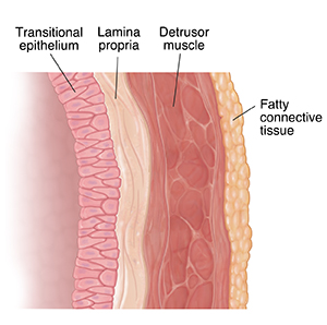
      The bladder wall is made of many layers, including:

      • Urothelium or transitional epithelium. This is the layer of cells that lines the inside of the kidneys, ureters, bladder, and urethra. Cells in this layer are called urothelial cells or transitional cells.

      • Lamina propria. This is the next layer around the urothelium. It’s a type of connective tissue.

      • Muscularis propria. This is the outer layer. It’s the thick muscle tissue outside the lamina propria.

      • Fatty connective tissue. This covers the outside of the bladder and separates it from other organs.

      Superficial bladder cancer affects only the inside lining of the bladder. This is the transitional epithelium. Cancer here is called transitional cell carcinoma (TCC) or urothelial carcinoma. As the cancer grows, it can become invasive bladder cancer that goes into deeper layers of the bladder wall. Over time, it can grow into the fatty connective tissue

      Related Topics

      • Neurological Exam for Children
      • Multiple Sclerosis (MS)

        nutch_noindex
        • About Us
        • Contact
        • MyChart Login
        • Careers
        • Blog
        • Refer a Patient
        • Download from App Store Download From Google Play
        • Notice of Nondiscrimination
        • Terms of Use
        • Privacy Policy
        • Code of Conduct

        © 2021 Stanford Children's Health

        About

        • About Us
        • Our Hospital
        • Patient Stories
        • Blog
        • News
        • Send a Greeting Card
        • New Hospital
        • Careers

        Connect

        • Donate
        • Refer a Patient
        • Contact Us
        • Pay Your Bill

        Find

        • Doctors
        • Locations
        • Services
        • Classes
        Download from App Store Download From Google Play

        Also Find Us on:

        • facebook
        • twitter
        • youtube
        • linkedin
        US News & World Report Badge
        • Stanford Medicine
        • Stanford University
        • Stanford Health Care
        • Lucile Packard Foundation
        • Notice of Nondiscrimination
        • Terms of Use
        • Privacy Policy
        • Code of Conduct
        • © 2021 Stanford Children's Health
        /nutch_noindex