Cholesteatoma
What is a Cholesteatoma?
A cholesteatoma is a skin cyst in the middle-ear made up of trapped skin cells and debris.
What causes Cholesteatoma?
Cholesteatomas can result from injury to the eardrum, chronic middle ear infections and/or chronic pressure buildup, which weakens the eardrum until a small pocket forms that stores trapped skin and debris. Pressure buildup can also be the result of a poorly functioning eustachian tube, the small opening that connects the middle ear to the nose and equalizes pressure in our ears. The trapped skin then continues to divide and grow, thus causing the cyst to grow. Some children can be born with a cholesteatoma when an accidental collection of skin is trapped behind the eardrum where no skin is expected to be.
Why is it a concern?
Cholesteatomas damage the eardrum, and they can also grow. As they grow, they can damage the neighboring structures of the middle ear and even the inner ear and/or brain if left untreated. They can dramatically affect and damage hearing, cause dizziness, and injury to facial nerves. Cholesteatomas can also cause infections in the middle ear that, left untreated, sometimes spread to the brain.
How do we evaluate it?
Evaluation for a cholesteatoma involves an examination of the eardrum by an Ear Nose and Throat specialist (Otolaryngologist). It is useful to have a formal hearing test, to assess the impact on hearing. Often, a CT scan, which uses a series of X-rays, is needed to further see the extent of the cholesteatoma. In certain cases, a special type of MRI scan called a diffusion weighted imaging MRI can also be helpful in detecting cholesteatoma.
How do we treat it?
Cholesteatomas are removed with surgery, which is typically done under general anesthesia. This surgery removes the cholesteatoma from the eardrum and the space behind the eardrum, called the middle ear and mastoid. It is often very difficult to remove all of the skin cells of the cholesteatoma in one surgery, therefore the process requires several procedures separated by months. Often, two or more surgeries are required for removal and surveillance to check for recurrence of the cholesteatoma, and to rebuild the hearing, if possible. At the time of surgery, anything damaged by the cholesteatoma can be repaired, such as the eardrum or the little hearing bones called the ossicles.
What is the long-term outlook of Cholesteatoma?
The long-term outlook for recovery is good. Hearing can be restored or aided with amplification after surgery depending on the extent of damage from the cholesteatoma. Infections can be treated with antibiotic ear drops or oral antibiotics as needed before and/or after surgery.
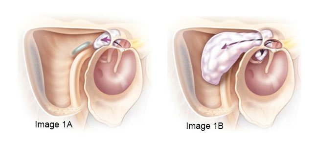 Cholesteatomas often start as a small skin cyst (Image 1A) in the upper part of the eardrum, but with time can grow into the mastoid, the bony airspace behind the ear canal (Image 1B), as well as lower to involve the hearing bones.
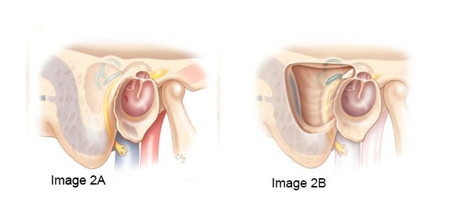 A view of a normal eardrum at the end of the ear canal (Image 2A) and the normal mastoid space behind the ear drum. The same view (Image 2B) after a mastoidectomy, which removes bone from the mastoid space in order to remove cholesteatoma.

Connect with us:
Download our App: