Ultrasound in Pregnancy
What is an ultrasound?
An ultrasound scan is a diagnostic technique which uses high-frequency sound
waves to create an image of the internal organs. A screening ultrasound is sometimes done during the course of a pregnancy to monitor normal fetal growth and verify the due date. Ultrasounds may be performed at various times throughout pregnancy for different reasons:
In the first trimester:
To establish the dates of a pregnancy
To determine the number of fetuses and identify placental structures
To diagnose an ectopic pregnancy or miscarriage
To examine the uterus and other pelvic anatomy
In some cases to detect fetal abnormalities
Mid-trimester: (sometimes called the 18 to 20 week scan)
To confirm pregnancy dates
To determine the number of fetuses and examine the placental structures
To assist in prenatal tests such as an amniocentesis
To examine the fetal anatomy for presence of abnormalities
To check the amount of amniotic fluid
To examine blood flow patterns
To observe fetal behavior and activity
To examine the placenta
To measure the length of the cervix
To monitor fetal growth
Third trimester:
To monitor fetal growth
To check the amount of amniotic fluid
As part of a biophysical profile
To determine the position of a fetus
To assess the placenta
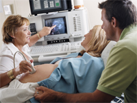
How is an ultrasound scan performed?
Although the specific details of each procedure vary slightly, generally, ultrasounds follow this process. Two types of ultrasounds can be performed during pregnancy:
Abdominal ultrasound. In an abdominal ultrasound, gel is applied to the abdomen and the ultrasound transducer glides over the gel on the abdomen to create the image. A woman may need to have a full bladder for abdominal ultrasounds in early pregnancy.
Transvaginal ultrasound. In a transvaginal ultrasound, a smaller ultrasound transducer is inserted into the vagina and rests against the back of the vagina to create an image. A transvaginal ultrasound produces a sharper image and is often used in early pregnancy.
There are several types of ultrasound imaging techniques. The most common is two dimensional, or 2D. This gives a flat picture of one aspect of the image.
If more information is needed, a 3D ultrasound examination can be performed. This technique, which provides a three-dimensional picture, requires a special machine and special training. But the 3D image allows the doctor to see width, height, and depth of images, which can be helpful in diagnosis. The 3D images can also be captured and saved for later review.
The latest technology is 4D ultrasound, which allows the health care provider to visualize the unborn baby moving in real-time. With 4D imaging, a three-dimensional image is continuously updated, providing a "live action" view. These images often have a golden color, which helps show shadows and highlights.
Ultrasound images may be captured in still photographs or on video to document findings.
What are the risks and benefits of ultrasound?
Fetal ultrasound has no known risks other than mild discomfort due to pressure from the transducer on the abdomen or in the vagina. No radiation is used during the procedure.
Transvaginal ultrasound requires covering the ultrasound transducer in a plastic or latex sheath, which may cause a reaction in patients with a latex allergy.
Fetal ultrasound is sometimes offered in nonmedical settings to provide keepsake images or videos for parents. While the ultrasound procedure itself is considered safe, it is possible that untrained personnel may give parents false assurances about their baby's well-being, or perhaps an abnormality may be missed. Having an ultrasound performed by trained medical personnel who can correctly interpret findings is recommended. Talk with your health care provider if you have questions.
Ultrasound is a technique that is constantly being improved and refined. As with any test, results may not be completely accurate. However, ultrasound can provide valuable information for parents and health care providers to help manage and care for the pregnancy and fetus. In addition, ultrasound gives parents a unique opportunity to see their baby before birth, helping them to bond and establish an early relationship.

Connect with us:
Download our App: