Notice of West building lobby closure at Lucile Packard Children’s Hospital Stanford

Sarah Dubner, MD
Clinical Associate Professor
Developmental Behavioral Pediatrics
Locations
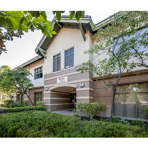

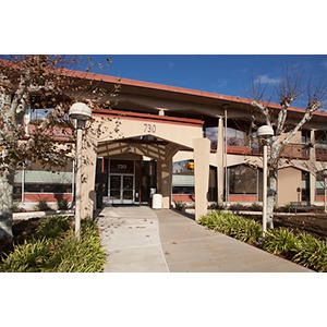
730 Welch Road
Palo Alto, CA 94304
Phone : (650) 497-8000
Fax : (650) 497-8034
2577 Samaritan Dr Ste 705
San Jose, CA 95124
Phone : (408) 523-3960
Fax : (650) 942-8348
Work and Education
Perelman School of Medicine University of Pennsylvania, Philadelphia, PA, 5/19/2008
University of Washington Pediatric Residency, Seattle, WA, 06/30/2011
Stanford University Developmental-Behavioral Pediatrics Fellowship, Palo Alto, CA, 07/06/2020
Developmental Behavioral Pediatrics, American Board of Pediatrics, 2021
Pediatrics, American Board of Pediatrics, 2011
Languages
English
Spanish

Connect with us:
Download our App: