Contact us
Contact us to learn more or if you are ready to schedule an appointment.
Notice of West building lobby closure at Lucile Packard Children’s Hospital Stanford
Congenitally corrected transposition of the great arteries (CCTGA) is a rare congenital heart defect. With CCTGA, the right and left ventricles are switched in position and in roles.
The normal heart has two muscular pumping chambers, called the right and left ventricles. These two ventricles are not identical in structure or function. They each have evolved and developed to serve very specific purposes. In a normal heart, the role of the left ventricle is to pump blood to the body. To do this, it must pump at high pressure. The left ventricle is designed for this role and performs it very well. On the other hand, the role of the right ventricle is to pump blood to the lungs. The right ventricle has a much easier task than the left ventricle, because it only has to pump blood at low pressure, typically about 15% of the pressure that the left ventricle uses. The right ventricle is designed to pump at this lower pressure, and it is structurally not suited to pump at higher pressure. In individuals with CCTGA, the ventricles are reversed and the stronger left ventricle pumps blood to the lungs while the weaker right ventricle has the harder task to pump blood to the entire body.
In the United States, only 0.5% to 1% of all babies born with heart defects have CCTGA. Most individuals born with CCTGA eventually need some type of heart surgery. There are several other heart abnormalities commonly found with CCTGA.
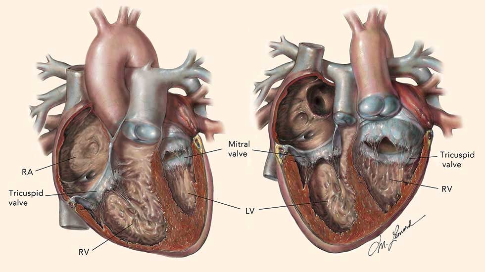
A normal heart, on the left, compared with a CCTGA heart on the right.
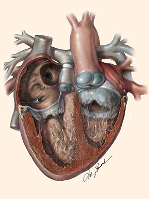
If the double-switch repair is the best option for you/your child, we may need to first strengthen the left ventricle by placing a pulmonary artery (PA) band, as shown in the center of this diagram. The band creates resistance to blood flow that makes the left ventricle work harder. This builds up the muscle of the left ventricle and its strength over time. In some cases, the PA band may need to be tightened. When the left ventricle becomes strong enough to pump blood to the body, we can then perform the modified double-switch repair.
Our center has developed five criteria that help us determine when a left ventricle is ready for double-switch surgery:
Congenitally corrected transposition of the great arteries (CCTGA) can occur alone or in conjunction with other forms of congenital heart disease—abnormalities in fetal heart development during pregnancy. Ninety percent of individuals with CCTGA also have other heart defects, such as a hole between the ventricles (a ventricular septal defect, or VSD), an abnormality of the tricuspid valve, or an obstruction in the pathway out of the left ventricle (left ventricular outflow tract obstruction with either pulmonary stenosis or pulmonary atresia). The following conditions are often associated with CCTGA:
Ventricular septal defect (VSD). In this common heart defect, there is a hole in the muscle wall that separates the left and right ventricles. Approximately 70% of all patients with CCTGA also have a VSD.
Pulmonary stenosis. The pulmonary valve of the heart connects the left ventricle to the pulmonary artery in CCTGA. When it doesn’t develop properly, it can limit blood flow. Approximately 60% of all patients with CCTGA also have pulmonary stenosis.
Pulmonary atresia. In some patients, the pulmonary valve does not form, and blood is not able to flow from the heart into the lungs. This is called pulmonary atresia.
Left ventricular outflow tract obstruction (LVOTO). This refers to a number of different types of pulmonary stenosis, or obstruction to blood flow as it leaves the left ventricle, common in CCTGA.
Ebstein’s anomaly of the tricuspid valve. This heart defect occurs when the tricuspid valve is abnormally formed. It often results in significant leakage or backflow of blood from the right ventricle into an upper chamber of the heart called the atrium (regurgitation). Approximately 20% of all patients with CCTGA also have Ebstein’s anomaly of the tricuspid valve.
Tricuspid regurgitation. When there is leakage of the tricuspid valve, which is the valve between the atrium and the right ventricle, blood flows backward into the atrium. It can occur in Ebstein’s anomaly but also can occur in tricuspid valves that have other abnormalities or appear structurally normal.
Dextrocardia. This refers to a condition where the heart is in the right side of the chest rather than the left, which is its normal location.
Heart block. This is an abnormal heart rhythm in which the electrical signals that generate the heartbeat are blocked between the upper and lower chambers of the heart, causing the heart to beat too slowly.
Contact us to learn more or if you are ready to schedule an appointment.

Connect with us:
Download our App: